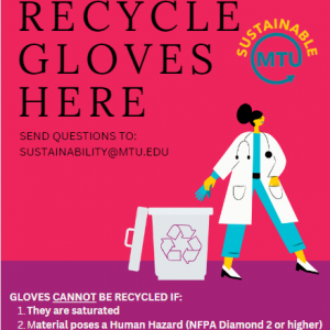
ACMAL recently installed new recycling infrastructure across the seven labs on the 6th and 7th floor of the M&M and the STEM lab at the ATDC.
Please review the new Recycling section in ACMAL Cleanliness under Policy.
ACMAL recently installed new recycling infrastructure across the seven labs on the 6th and 7th floor of the M&M and the STEM lab at the ATDC.
Please review the new Recycling section in ACMAL Cleanliness under Policy.
Research Scientist, Materials Science and Engineering
Michigan Technological University
Materials Science and Engineering Seminar
Thursday, April 13, 2023, 1 – 2 p.m.
Minerals and Materials Engineering Building (M&M) , 610
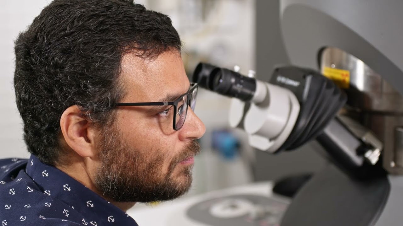
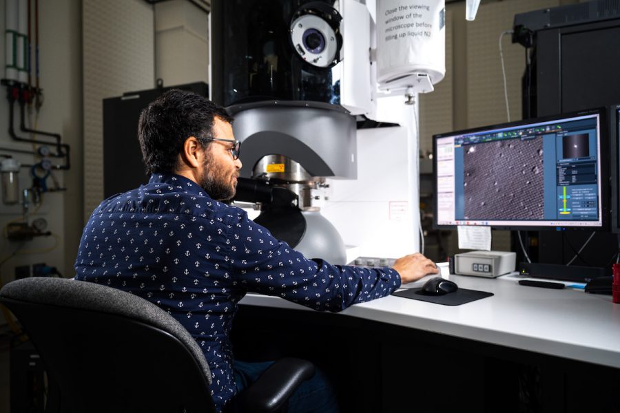
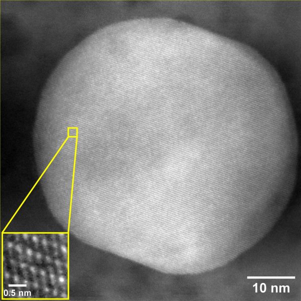
Tiny things lead to big discoveries in the Electron Optics Facility that houses Titan, the ultra-sensitive microscope that analyzes materials at the atomic level. The Titan Themis Scanning Transmission Microscope, or S-TEM, has its own dedicated and stabilized space, complete with water-cooled temperature controls and back-up power. A powerful tool in an extensive array of electron microanalytical and X-ray instruments in Michigan Tech’s Applied Chemical and Morphological Analysis Laboratory (ACMAL), Titan is one of only two microscopes of its kind in the state of Michigan. In addition to high-resolution images, Titan can also perform fractional or chemical analysis. Its applications are useful in many research areas, including health, industrial, and technology. The electron optics facility is managed by Erico Freitas, a research scientist who runs the majority of samples but also trains students on how to use the equipment.
Are you a:
Are you planning to seek funds for research elsewhere, but require start-up money to develop your proposal?
If so, you are eligible to apply for Seedling Research Funding opportunities of up to $1,000.
The Applied Chemical and Morphological Analysis Laboratory (ACMAL) at Michigan Tech has set aside $4,000 to fund pre-proposal research. These funds are designed to be granted to those who need to do preliminary investigations and data collection, which will then be used to develop a proposal for research funding from another source.
We would like to officially welcome our two new lab assistants: Zoe Hoffman and Maci Dostaler. Also, congratulations to our previous senior lab assistant, Aleister Kerr. Aleister has taken a new position off campus and will no longer be working with us.
Zoe Hoffman is a current student at Michigan Tech majoring in Medical Laboratory Science with a minor in Pre-Health from southeast Michigan. She has always found the idea of working in a laboratory fun and likes to assist in the ACMAL unit.

Maci Dostaler is currently an undergraduate student in Michigan Tech’s Biomedical Engineering program. She has lived in the Copper Country all of her life and is a proud first-generation college student. After graduation, she is hoping to work with imaging and non-invasive lasers. She is involved deeply around campus from volunteering to Greek life! In her free time she loves to challenge herself in snowboarding competitions both throughout the midwest and nationally.
You can contact Maci and Zoe if you need assistance with sample preparation or coating.
Dr. Edward Laitila is a kind and humble science teacher who cannot be appreciated enough for his dedication to the advancement of science and the education of students. Over the years he has developed methods of analysis and processes for material development that are unique, practical, and yield powerful results. His way of teaching takes extra time to convey not only knowledge, but to impart skills in critical thinking that are too-often absent from a regular education. For his astounding work over many years, it is a pleasure to state just how valuable an asset he has been to Michigan Tech, to students, and to the scientific community.
Professor Laitila teaches courses in crystallography, diffraction, and materials forensics within the Materials Science and Engineering department. His official titles and positions are Senior Research Engineer/Scientist II and Adjunct Assistant Professor. His main duties are to act as the manager and sole employee of the X-Ray Facility, which operates under the Applied Chemical and Morphological Analysis Laboratory (ACMAL). There, he manages, operates and repairs the various X-Ray Diffraction (XRD) and X-Ray Fluorescence (XRF) equipment.
Dr. Laitila started running the X-Ray Facility in 1983. At that time he had a two-year electrical engineering degree. He then obtained a bachelor’s degree and spent the next 20 years researching and working. Eventually he found extra time to write a report on his years of research and to finish his PhD. He has many undergrad and graduate students doing research for him. He also teaches classes and helps students with planning their own research experiments and writing research papers. He now has nearly 40 years of experience in XRD.
XRD is a process of metrology which characterizes a material by measuring the size of, and pattern to, its atomic structure. X-rays are waves, and crystalline materials are made up of periodic arrangements of atoms in a crystal lattice structure. Every crystalline material has a pattern that is unique, referred to as a diffraction fingerprint. XRD involves wave behavior similar to light diffraction, such as that seen in the double slit experiment. The x-ray waves travel through spacing and can constructively or destructively interfere with each other, which results in the unique pattern. This produces a pattern of peaks and dark sections which can be analyzed to determine the material structure and the distance between the atoms themselves. Through XRD researchers can obtain pinpoint accuracy of a material’s lattice structure, chemical composition, and phase composition.
Dr. Laitila’s research interests are X-ray diffraction theory and its many applications, including mechanical alloying, intermetallic materials, physical metallurgy, nanoscale materials, materials characterization, additive manufacturing, and powder metallurgy. His latest published research was featured in the journal Powder Diffraction, Vol. 23, Issue 2 and was entitled “Employing X-ray scattering to characterize materials with grain sizes in the nano-regime.” In it he explained his creation of a new analytical technique for measuring the number of atoms in the grain boundaries of a nanoscale material. He was chosen for publishing after giving a talk at an annual X-Ray powder diffraction conference. On that work, he remarked, “Even though I did it on one material, I showed that this could be applied to anything.” From that research he is now working on a process which can make iron directly from iron ore without a need for using carbon. Such a process is exciting because it could revolutionize the steel industry in a way that is net positive for earth’s climate.
In recent research Dr. Laitila has come up with a process for making nano-composites. With his novel method it is simple to vary the amount of a second phase by varying the milling time. He has dubbed it Mechanonanosynthesis. He explained that it will be ideal in the creation of powders for additive manufacturing.
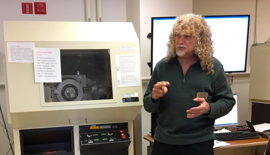
Dr. Laitila went into detail about some interesting projects he has done over the years. He explained that in 1983 the diffractometers were all analog machines, and that only a couple of types had been automated. The lab worked with a teletype, which put little holes on tape that would collect the data. Researchers would then put those in a mainframe computer to do analysis. He was asked in ‘84 if he could interface the diffractometer with an early-model PC, which had an 8088 processor. He used Basic to code an MS-DOS program for the interface. He laughed as he recalled the story, saying, “I got it to work, and got it to collect data, and then we bought an automated system.”
The professor describes himself as a big proponent of critical thinking. He explained, “I honestly believe we have got a major problem in our education system, because we teach knowledge instead of teaching how to think. Teachers here have critical thinking skills but we don’t usually focus on that. When a student comes to me with a question I try to return with a question that forces them to think of a way to answer their own question. Usually they have the knowledge but they don’t know how to piece it together. I try to piece things together from different subjects and how those things combine in material science. I try to take every opportunity to teach.” He explained that his favorite part of teaching is “seeing the light go on when a student gets the subject.”
In addition to all his other duties, Dr. Laitila spends extra time teaching teenagers about science. He has run a session, as part of the Women in Engineering and Engineering Scholars program, for many years. Each summer for more than 50 years, Michigan Tech Summer Youth Programs (SYP) has welcomed to campus more than 1,000 youth from grades 6–11, from across the country and around the world. SYP students come for week-long, hands-on, experiential learning in one or more of their 50+ week-long explorations in science, technology, engineering, and mathematics (STEM), humanities, and law. Dr. Ed is a favorite among many students, especially those in the SYP. Last year, when students were finishing SYP and were asked what program was their favorite, they chanted Ed’s name.
Dr. Edward Laitila is a catalyst for the process of science. He is a man that makes the magic happen. He is a student favorite, an expert in XRD, and a valuable researcher. Anyone interested in doing work with Edward or using the equipment in his lab should contact him or get in touch with ACMAL Director Elizabeth Miller for more information. Remote teaching and research are available. If you are a student with a project that requires XRD or are interested in helping Dr. Laitila with his research, there may be opportunities available to you.
There is no better place to get involved in some exciting research!
By Joshua Jongema.
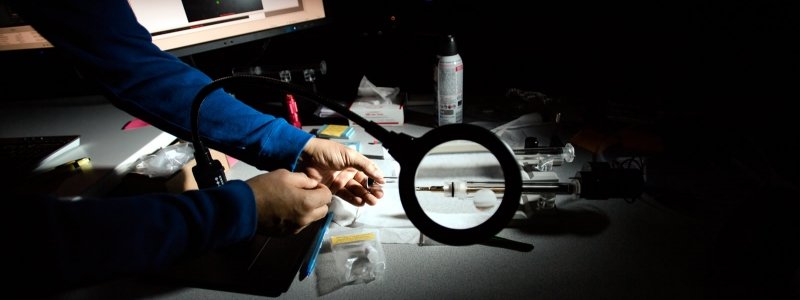
A materials science and engineering team of students Sonja Blickley, Tori Nizzi, Anna Palmcook, and Austin Schaub were sponsored by Hobart Brothers LLC. (Hobartbrothers.com) to develop a new process that has yielded some exciting results. Working with Dr. Erico Freitas, operator of the Titan Themis Scanning Electron Microscope, these students used the FEI 200kV Titan Themis Scanning Transmission Electron Microscope (STEM) to produce some awesome images of a welded material. They have granted special permission to show these pictures here, despite wishing to keep their work and the composition of their material confidential.
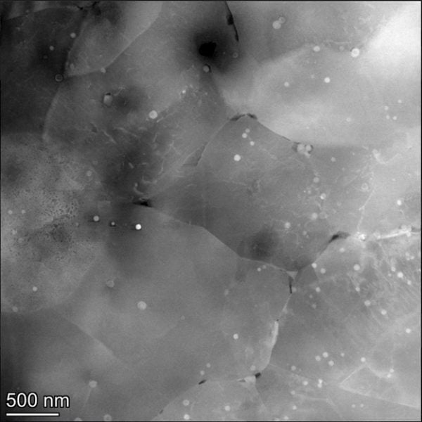
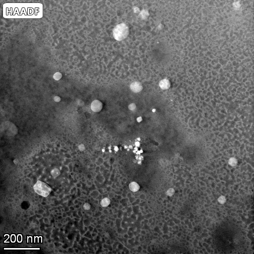
This team is very excited about their results, which help to drive the science of materials and engineering forward. Congratulations to them for their hard work paying off!
By Joshua Jongema.
Good morning,
This is just a reminder that the ACMAL facilities will be CLOSED Thursday and Friday, Nov. 24th and 25th, 2022, in observance of Thanksgiving. This is a university holiday so the building will also be locked. Only those with swipe access will be able to use the instruments. Please plan your instrument use accordingly.
I will be on vacation starting at noon tomorrow returning to work on Monday 11/28. Dr. Freitas, Dr. Leftwich, and Mr. Josh King will be available through Wednesday (11/23). Dr. Laitila will be available through Tuesday (11/22).
In case of an emergency call public safety.
Happy Holidays,
Liz
Department of Biological Sciences Seminar Series
Research Scientist
Materials Science and Engineering
Michigan Technological University
Dr. Freitas manages the FEI 200kV Titan Themis STEM in the Electron Optics Facility of the Applied Chemical and Morphological Analysis Laboratory (ACMAL) at Michigan Tech. Transmission electron microscopy (TEM) is widely used for morphological studies of biological systems as it offers a much higher spatial resolution (over 200 times) than optical microscopy. During this seminar Dr. Freitas will present the capabilities of the Scanning transmission electron microscope laboratory at MTU and will address the types of ex-situ and in-situ analysis that can be conducted for biological systems using a diversity of approaches available through the ACMAL.
For additional information, visit the Department of Biological Sciences.
Thursday, September 29, 2022
3–4 p.m. GLRC 202
If you had plans to use the STEM in the August-September I’ll be unavailable on the following dates:
August 1st – 5th (out of town); returning flight schedule for Friday 5th in the morning I’ll be available for online.
August 18th – September 7th (out of town)
Regards, Erico
Erico Freitas
Research Scientist
Applied Chemical and Morphological Analysis Laboratory
Materials Science and Engineering
Michigan Technological University
1400 Townsend Drive
Houghton, MI 49931-1295
Office: Room 622 Minerals & Materials Building
(906) 299-2714 or efragafr@mtu.edu