Author: College of Engineering

Sue Hill is the Digital Content Manager for the College of Engineering.

STEM Titan Themis unavailable from July 18-22

I would like to let you know I’ll be on vacation from July 18th to 22nd and will not be available to run TEM sessions or training sessions during that period of time at the FEI 200kV Titan Themis STEM.

Also, a new rooftop chiller will be installed soon at the STEM lab and the instrument will be unavailable. I’ll let you know when that is going to happen.

Regards,

Erico Freitas 
Who is Studying the Failure Mechanisms of Electrical Wire Terminals at Michigan Tech?

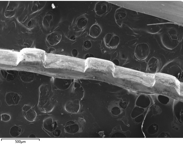
Micrograph of a wire on a substrate with a 500 micron scale marker.

An Advanced Metalworks Enterprise undergraduate student team, sponsored by Lear Corporation, is studying the performance of copper electrical wires in automobiles. Corrosion is the most common failure mechanism of wires used in crimp connectors; deformation in the wire terminal’s tin plating can cause additional contact issues within the connector. Electron microscopy aids in pinpointing the location of corrosion products on the wire and observing deformation in the tin plating. With this analysis, the team can now explore ways to improve the wire quality or crimping mechanism to minimize wire failures.

Image taken by Eli Harma and Reese Eichner, senior undergraduate materials science and engineering students, on Philips XL 40 ESEM.

Learn more about the Advanced Metalworks Enterprise at MTU: AME Website

Visit the Applied Chemical and Morphological Analysis Laboratory’s webpage to learn more about our shared facility and instruments available to the Michigan Tech research community: ACMAL

Who is Studying Miniature Magneto-optic Devices at Michigan Tech?

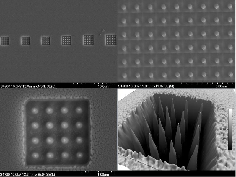
Four images on different scales showing pillar arrays on a surface.

The successful fabrication of miniature optical components is key for progressing current optical technologies. A family of such miniature optical components must be able to efficiently rotate linearly polarized light at small scales. Estefanio Kesto, under the guidance of Dr. Miguel Levy, is studying the interaction between light and ferromagnetic iron garnet nanostructures. It has been observed that the polarization rotation of linearly polarized light, known as the magneto-optic response, traveling through such a nanostructure will be enhanced. The ferromagnetic iron garnet nanostructures pictured above, which enhance the magneto-optic response, are being studied to further miniaturize polarization rotators and other interferometric components. Additionally, Professor Levy and his research group are diving into the unexplored region of magneto-optic beam splitting and its applications in classical and quantum computing.

Pillars fabricated and image taken by Estefanio Kesto, undergraduate student in electrical engineering, using ACMAL’s Hitachi FB-2000A FIB, Hitachi S-4700 FE-SEM, and Asylum Research MFP-3D Origin+ AFM.

Read more about the Dr. Miguel Levy’s research in the following articles:

All-dielectric magnetic metasurface for advanced light control in dual polarizations combined with high-Q resonances

Nonreciprocal magneto-optic beam splitting

Two-dimensional array of iron-garnet nanocylinders supporting localized and lattice modes for the broadband boosted magneto-optics

Visit the Applied Chemical and Morphological Analysis Laboratory’s webpage to learn more about our shared facility and instruments available to the Michigan Tech research community: ACMAL

ACMAL Welcomes Erico T. F. Freitas

Erico T. F. Freitas

Research Scientist Erico T. F. Freitas has joined ACMAL and Materials Science and Engineering. He will manage the FEI 200kV Titan Themis STEM in ACMAL’s Electron Optics Facility. Freitas has experience in nanostructured materials characterization, with specialties in TEM, STEM, EELS, EFTEM, EDX, and electron diffraction.

Freitas worked previously in the multiuser electron microscopy facility managing the TEM at the Universidade Federal de Minas Gerais, Brazil.

ACMAL Holiday Schedule

ACMAL will have limited hours over the upcoming holiday break. 
The university will be closed on Dec 23 through Jan 1.  The building will be locked on those days. Unrestricted users will have unlimited access to the building and labs.  Restricted users have access during the other days.  We ask that newly trained users make appointments during the period so someone is near in case there are troubles.

Dr. Ed Laitila and Liz Miller will be out the entire 2 week period.  Dr. Tim Leftwich, Josh King, Kyle Hrubecky, and Aleister Kerr will be available by appointment.

If there are problems contact us.  Our contact information is:

Liz: 906-370-6538
Ed: 906-369-2041
Aleister: 520-576-3557
Josh: 404-808-2803
Tim: trleftwi@mtu.edu

Happy Holidays,

Elizabeth Miller

Who is Imaging Hemolysin X Treated Red Blood Cells at Michigan Tech?

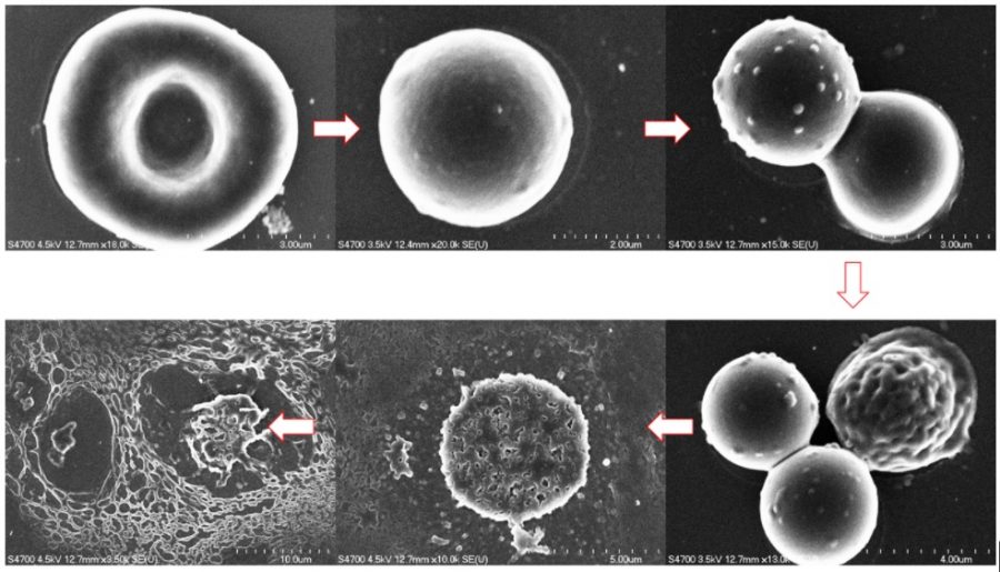
Sequence of six images showing the disintegration of cell membranes.

The Laboratory of Mechanistic Glycobiology research group, led by Dr. Tarun Dam, is studying how the function of biomolecules from plant cells translates to human cells. Hemolysin X is a biomolecule that can disrupt and disintegrate cell membranes. The image above depicts how Hemolysin X systematically disintegrates a red blood cell.  The research group is looking into how this molecule reacts with other types of mammalian cells, including cancer cells.

Image taken by Jared Edwards, Chemistry PhD candidate, on ACMAL’s Hitachi S-4700 FE-SEM.

Learn more about the Laboratory of Mechanistic Glycobiology research group: Laboratory of Mechanistic Glycobiology

Visit the Applied Chemical and Morphological Analysis Laboratory’s webpage to learn more about our shared facility and instruments available to the Michigan Tech research community: ACMAL

Who is Imaging Electrospun Polycaprolactone Fiber Scaffolding at Michigan Tech?

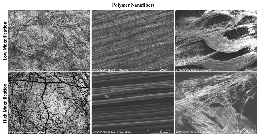
Six panels of three different polymer nanofibers at low and high magnifications.

Dr. Smitha Rao, assistant professor for Biomedical Engineering at Michigan Tech, and the Biomedical µDevices research team developed a way to be able to observe how breast cancer cells grow and migrate in various environments. The project developed scaffolding systems that mimic structures that could be found in human tissue. They engineered three polycaprolactone scaffold structures to test different topographical and mechanical features: hexagonal, mesh-like and aligned.

The image was taken by Dr. Smitha Rao’s graduate and undergraduate students using ACMAL’s Hitachi S-4700 FE-SEM.

Read more about Dr. Rao and the Biomedical µDevices research team’s work:

Visit the Applied Chemical and Morphological Analysis Laboratory’s webpage to learn more about our shared facility and instruments available to the Michigan Tech research community: ACMAL

If you have you have an image you would like to be featured, submit it here: