Category: Electron Optics Facility Featured

Researchers Contribute to the Database on the Magnetic Properties of Meteorites

Cover of Icarus

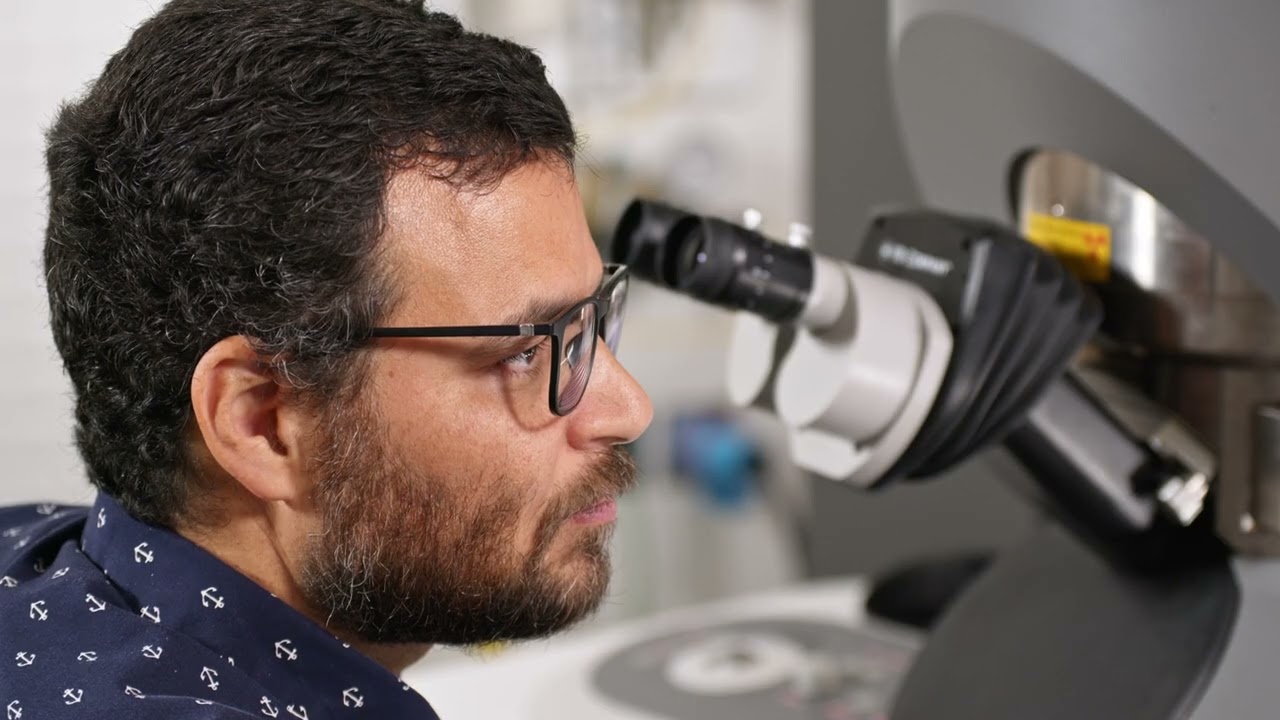
Aleksey Smirnov (GMES) and Katie Bristol ’20 (M.S. Geophysics), currently a Ph.D. student at the University of Florida, collaborated as co-authors on a research paper published in Icarus, a prestigious journal renowned in the field of planetary science.

The paper is titled “Magnetic characterization of the Daule chondrite (Ecuador’s first meteorite fall): The case of elusive tetrataenite?”

The study also involved researchers hailing from Ecuador, Iceland and Norway.

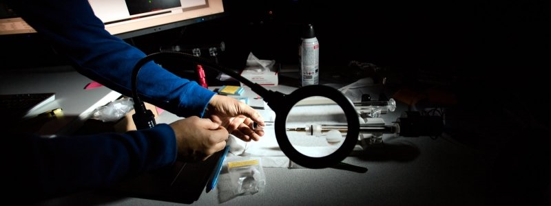
Two representative samples of bulk Daule material were analyzed using an FEI Philips XL 40 Environmental Scanning Electron Microscope (ESEM) at the Applied Chemical and Morphological Analysis Laboratory at Michigan Technological University (MTU).

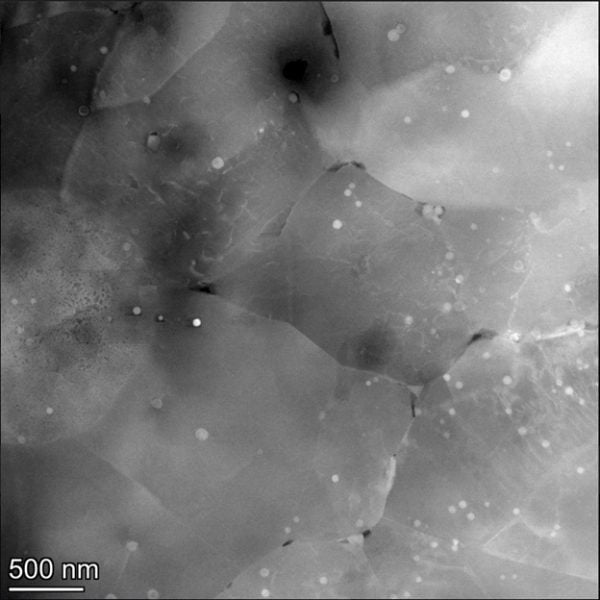
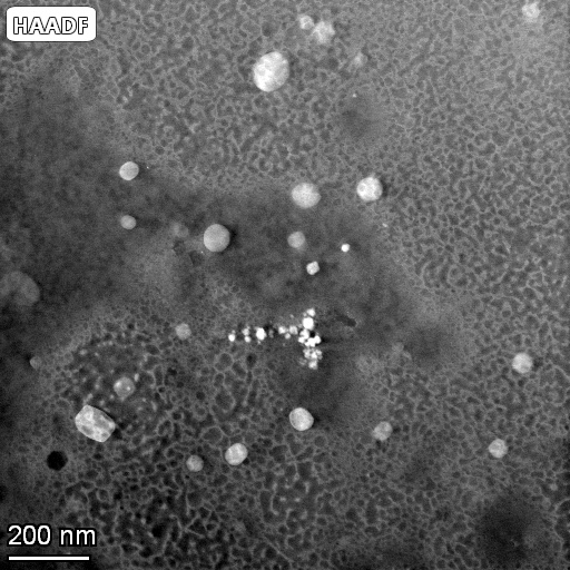
  • The morphology and texture of mineral grains were examined using backscattered electron (BSE) imaging.
  • The elemental composition of the material was determined through energy-dispersive x-ray spectroscopy (EDS) using a ∼ 15 kV accelerating voltage.
  • Basic elemental mapping was performed.

The new data contribute to the database on the magnetic properties of meteorites, in which meteorites of Daule’s type are underrepresented.

K.E. Bristol, A.V. Smirnov, E.J. Piispa, M.R. Ramirez Navas, A. Kosterov, E.V. Kulakov, Magnetic characterization of the Daule chondrite (Ecuador’s first meteorite fall): The case of elusive tetrataenite?, Icarus, Volume 404, 2023.

https://doi.org/10.1016/j.icarus.2023.115684

Under the Titan Lens: Microscope Takes Research to Atomic-Level

Play Under the Titan Lens: Microscope Takes Research to Atomic-Level video
Preview image for Under the Titan Lens: Microscope Takes Research to Atomic-Level video

Under the Titan Lens: Microscope Takes Research to Atomic-Level

Tiny things lead to big discoveries in the Electron Optics Facility that houses Titan, the ultra-sensitive microscope that analyzes materials at the atomic level. The Titan Themis Scanning Transmission Microscope, or S-TEM, has its own dedicated and stabilized space, complete with water-cooled temperature controls and back-up power. A powerful tool in an extensive array of electron microanalytical and X-ray instruments in Michigan Tech’s Applied Chemical and Morphological Analysis Laboratory (ACMAL), Titan is one of only two microscopes of its kind in the state of Michigan. In addition to high-resolution images, Titan can also perform fractional or chemical analysis. Its applications are useful in many research areas, including health, industrial, and technology. The electron optics facility is managed by Erico Freitas, a research scientist who runs the majority of samples but also trains students on how to use the equipment.

Funding Announcement: Seedling Research

Sample preparation on a benchtop.

Planting the Seeds of Research

Are you a:

  • Tenured, tenure-track, or research faculty?
  • Post-doctoral researcher?
  • Michigan Tech staff member?

Are you planning to seek funds for research elsewhere, but require start-up money to develop your proposal?

If so, you are eligible to apply for Seedling Research Funding opportunities of up to $1,000.

The Applied Chemical and Morphological Analysis Laboratory (ACMAL) at Michigan Tech has set aside $4,000 to fund pre-proposal research. These funds are designed to be granted to those who need to do preliminary investigations and data collection, which will then be used to develop a proposal for research funding from another source.

Learn More and Apply

Student Team Shares Exciting Images

A materials science and engineering team of students Sonja Blickley, Tori Nizzi, Anna Palmcook, and Austin Schaub were sponsored by Hobart Brothers LLC. (Hobartbrothers.com) to develop a new process that has yielded some exciting results. Working with Dr. Erico Freitas, operator of the Titan Themis Scanning Electron Microscope, these students used the FEI 200kV Titan Themis Scanning Transmission Electron Microscope (STEM) to produce some awesome images of a welded material. They have granted special permission to show these pictures here, despite wishing to keep their work and the composition of their material confidential.

Micrograph with 500 nm scale showing dotted features on larger grains with boundaries.
Image 1 – This image shows a dispersed material within a matrix on the nanometer scale.
Micrograph on the 200 nm scale showing some of the dot features and the texture of the material.
Image 2 – This image is also a section of the first, zoomed in 70,000 times! To achieve this resolution the team used a High-Angle Annular Dark Field (HAADF) imaging technique.
Micrograph at the 10 nm of the dot feature, with a zoom showing its atomic structure.
Image 3 – This image is a section of the first, zoomed in 1,600,000 times! This was also taken using HAADF.

This team is very excited about their results, which help to drive the science of materials and engineering forward. Congratulations to them for their hard work paying off!

By Joshua Jongema.

Who is Studying the Failure Mechanisms of Electrical Wire Terminals at Michigan Tech?

Micrograph of a wire on a substrate with a 500 micron scale marker.

An Advanced Metalworks Enterprise undergraduate student team, sponsored by Lear Corporation, is studying the performance of copper electrical wires in automobiles. Corrosion is the most common failure mechanism of wires used in crimp connectors; deformation in the wire terminal’s tin plating can cause additional contact issues within the connector. Electron microscopy aids in pinpointing the location of corrosion products on the wire and observing deformation in the tin plating. With this analysis, the team can now explore ways to improve the wire quality or crimping mechanism to minimize wire failures.

Image taken by Eli Harma and Reese Eichner, senior undergraduate materials science and engineering students, on Philips XL 40 ESEM.

Learn more about the Advanced Metalworks Enterprise at MTU: AME Website

Visit the Applied Chemical and Morphological Analysis Laboratory’s webpage to learn more about our shared facility and instruments available to the Michigan Tech research community: ACMAL

Who is Studying Miniature Magneto-optic Devices at Michigan Tech?

Four images on different scales showing pillar arrays on a surface.

The successful fabrication of miniature optical components is key for progressing current optical technologies. A family of such miniature optical components must be able to efficiently rotate linearly polarized light at small scales. Estefanio Kesto, under the guidance of Dr. Miguel Levy, is studying the interaction between light and ferromagnetic iron garnet nanostructures. It has been observed that the polarization rotation of linearly polarized light, known as the magneto-optic response, traveling through such a nanostructure will be enhanced. The ferromagnetic iron garnet nanostructures pictured above, which enhance the magneto-optic response, are being studied to further miniaturize polarization rotators and other interferometric components. Additionally, Professor Levy and his research group are diving into the unexplored region of magneto-optic beam splitting and its applications in classical and quantum computing.

Pillars fabricated and image taken by Estefanio Kesto, undergraduate student in electrical engineering, using ACMAL’s Hitachi FB-2000A FIB, Hitachi S-4700 FE-SEM, and Asylum Research MFP-3D Origin+ AFM.

Read more about the Dr. Miguel Levy’s research in the following articles:

All-dielectric magnetic metasurface for advanced light control in dual polarizations combined with high-Q resonances

Nonreciprocal magneto-optic beam splitting

Two-dimensional array of iron-garnet nanocylinders supporting localized and lattice modes for the broadband boosted magneto-optics

Visit the Applied Chemical and Morphological Analysis Laboratory’s webpage to learn more about our shared facility and instruments available to the Michigan Tech research community: ACMAL

Who is Imaging Hemolysin X Treated Red Blood Cells at Michigan Tech?

Sequence of six images showing the disintegration of cell membranes.

The Laboratory of Mechanistic Glycobiology research group, led by Dr. Tarun Dam, is studying how the function of biomolecules from plant cells translates to human cells. Hemolysin X is a biomolecule that can disrupt and disintegrate cell membranes. The image above depicts how Hemolysin X systematically disintegrates a red blood cell.  The research group is looking into how this molecule reacts with other types of mammalian cells, including cancer cells.

Image taken by Jared Edwards, Chemistry PhD candidate, on ACMAL’s Hitachi S-4700 FE-SEM.

Learn more about the Laboratory of Mechanistic Glycobiology research group: Laboratory of Mechanistic Glycobiology

Visit the Applied Chemical and Morphological Analysis Laboratory’s webpage to learn more about our shared facility and instruments available to the Michigan Tech research community: ACMAL

Who is Imaging Electrospun Polycaprolactone Fiber Scaffolding at Michigan Tech?

Six panels of three different polymer nanofibers at low and high magnifications.

Dr. Smitha Rao, assistant professor for Biomedical Engineering at Michigan Tech, and the Biomedical µDevices research team developed a way to be able to observe how breast cancer cells grow and migrate in various environments. The project developed scaffolding systems that mimic structures that could be found in human tissue. They engineered three polycaprolactone scaffold structures to test different topographical and mechanical features: hexagonal, mesh-like and aligned.

The image was taken by Dr. Smitha Rao’s graduate and undergraduate students using ACMAL’s Hitachi S-4700 FE-SEM.

Read more about Dr. Rao and the Biomedical µDevices research team’s work:

Visit the Applied Chemical and Morphological Analysis Laboratory’s webpage to learn more about our shared facility and instruments available to the Michigan Tech research community: ACMAL

If you have you have an image you would like to be featured, submit it here:

Spontaneous Selective Deposition of Iron Oxide Nanoparticles on Graphite as Model Catalysts

Defect generation process steps.

Chathura de Alwis (Chemistry), Timothy R. Leftwich (MSE), Pinaki Mukherjee (MSE), Alex Denofrea (Chemistry) and Kathryn A. Perrine (Chemistry) published a paper titled “Spontaneous selective deposition of iron oxide nanoparticles on graphite as model catalysts” in Nanoscale Advances in 2019.

DOI: 10.1039/c9na00472f

Extract

Iron oxide nanomaterials participate in redox processes that give them ideal properties for their use as earth-abundant catalysts. Fabricating nanocatalysts for such applications requires detailed knowledge of the deposition and growth. We report the spontaneous deposition of iron oxide nanoparticles on HOPG in defect areas and on step edges from a metal precursor solution.

Various defects were created on the highly oriented pyrolytic graphite (HOPG) surface using either argon (Ar+) sputtering or a focused ion beam (FIB) to provide defects for nucleation sites. A Hitachi 2000 A FIB instrument was used to create tailored arrays of defects on HOPG using a Ga+ beam.

The sputter rate was calculated using the amount of materials removed, by recording a height profile of 1 nm using atomic force microscopy (AFM) and the time to sputter the pattern.

All the samples were imaged using a Hitachi S-4700 cold field emission high resolution field emission scanning electron microscopy (FE-SEM) instrument.

X-ray photoelectron spectroscopy (XPS) was performed using a PHI 5800 to analyze the elemental composition and oxidation state of surface species of the iron oxide nanoparticles grown on the HOPG surface.

Scanning transmission electron microscopy (STEM) imaging and energy dispersive X-ray spectroscopy (EDS) mapping were used to measure the phase and composition of iron oxide nanoparticles after annealing and to confirm if the deposition was preferential at the defect sites of graphite. A FEI Titan Themis aberration corrected scanning transmission electron microscope was used to obtain atomically resolved electron images and EDS maps of the iron oxide nanoparticles on the graphene coated TEM grid. The microscope was operated at 200 kV using a point resolution of the aberration corrected STEM mode of 0.08 nm. The microscope was equipped with a SuperX™ X-ray detector, which is composed of 4 detectors for fast X-ray mapping in STEM mode. The EDS mapping of the sample was performed on specific particles with an average beam current of 100 pA.

Acknowledgements

Equipment for obtaining the AFM images in this project was provided by NSF CHE #1725818. The electron microscopy research was performed at the Applied Chemical and Morphological Analysis Laboratory, at Michigan Technological University. The electron microscopy facility is supported by NSF MRI 1429232. We acknowledge the Michigan Tech REF-RS fund for support of this work and the David J. and Valeria Pruett Graduate Research Fellowship. We acknowledge the Applied Chemical and Morphological Analysis Laboratory (ACMAL) for staff assistance and use of facilities.

Recommended Citation

de Alwis, C., Leftwich, T., Mukherjee, P., Denofre, A., & Perrine, K. (2019). Spontaneous selective deposition of iron oxide nanoparticles on graphite as model catalysts. Nanoscale Advances, 1(12), 4729-4744.

http://doi.org/10.1039/C9NA00472F

Retrieved from: https://digitalcommons.mtu.edu/michigantech-p/1246

FESEM and FIB Used in Area-selective Atomic Layer Deposition Research

Surface Science cover for volume 690 December 2019.
Raman confocal images of Al2O3 make the cover of Surface Science.

Graduate students Mikhail Trought (Chemistry) and Chathura de Alwis (Chemistry), with undergraduate student alumnus Isobel Wentworth (ChemEng), research assistant professor Timothy R. Leftwich (MSE), and assistant professor Kathryn A. Perrine (Chemistry) published a paper titled “Influence of surface etching and oxidation on the morphological growth of Al2O3 by ALD” in Surface Science on August 9, 2019.

https://doi.org/10.1016/j.susc.2019.121479

The authors acknowledge the Applied Chemical & Morphological Analysis Laboratory (ACMAL) at Michigan Technological University for use of instruments and staff assistance, including Director Owen Mills, for training on the FESEM and FIB.

M. Trought and K. Perrine prepared the samples at Michigan Tech and at the Univ. of Minnesota, performed the surface analysis, analyzed all data collected, and wrote the manuscript. T. Leftwich assisted with the XPS data collection and analysis, and reviewing & editing the manuscript. I. Wentworth and C. de Alwis assisted with sample preparation and FTIR analysis. K. Perrine conceptualized the project.