Tag: csa

College of Sciences and Arts

Keyence VHX & LHX Lunch and Learn March 20, 2024

Detail from a Keyence microscope illustration.

I am pleased to extend an invitation to you for a Lunch and Learn session with Keyence on March 20th, from 10 a.m. to 12 p.m.

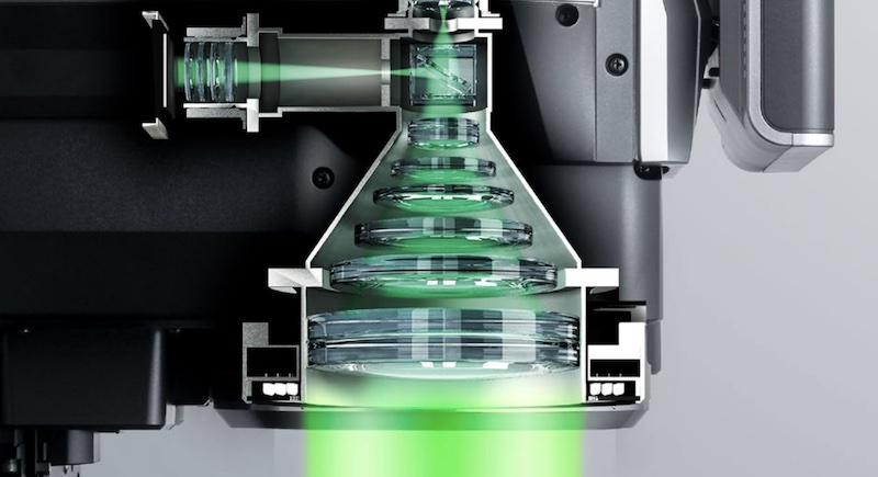
Keyence is a leading provider of digital microscopes, and this session will provide valuable insights into their cutting-edge technologies and products. It’s a fantastic opportunity to learn about the latest advancements in the field and discover how Keyence’s solutions can benefit your research.

The event will include presentations, demonstrations, and interactive discussions, followed by a complimentary lunch.

Date: March 20th
Time: 10 a.m. to 12 p.m.
Location: M&M 610

Please RSVP by March 15 using the RSVP FORM to confirm your attendance. Feel free to share this opportunity with any colleagues or members of your research group who may be interested. We look forward to having you join us for this informative session.

RSVP Form

Best,
Liz

Elizabeth Miller

ESEM Upgrade to SEMTech 8000

Instrument Offline from 8/1 to 8/18

SEMView8000 Operator Console

I am writing to inform you about an exciting upgrade to our Environmental Scanning Electron Microscope (ESEM) system. ACMAL has received funding to upgrade the ESEM with the SEMView8000. We would like to take this opportunity to thank the Vice President for Research Office, the Institute for Materials Processing, the Materials Science and Engineering Department, and the MSE Senior Design team for making this upgrade possible.

The instrument will be temporarily offline from August 1 to August 18 to facilitate the installation of the SEMTech 8000 upgrade. During the upgrade period, the ESEM system will be inaccessible, and no reservations or usage will be possible. We kindly request that you plan your projects accordingly and make alternative arrangements for your imaging and analysis requirements during this period.

To ensure a smooth transition and help you familiarize yourself with the upgraded system, we will be offering training sessions for all ESEM users. These training sessions are mandatory for anyone intending to use the SEMTech 8000. The sessions will be conducted starting from August 19, once the upgrade is complete.

To schedule your training session, please sign up for training. You will be directed to our self-scheduling calendar, where you can choose a time slot that suits you best. Appointments are available starting August 19. Due to limited availability, we recommend scheduling your session at your earliest convenience.

We understand that this temporary unavailability and the training requirement may cause some inconvenience, and we apologize for any disruption it may cause to your work. However, we believe that the benefits of the SEMTech 8000 upgrade and the subsequent training will be instrumental in advancing our research capabilities.

If you have any urgent projects or time-sensitive samples requiring ESEM imaging during the upgrade period, please let us know as soon as possible. We will do our best to accommodate your needs either before or after the downtime.

We appreciate your understanding and patience during this upgrade process. The improved capabilities of the SEMTech 8000, along with the training sessions, will undoubtedly enhance our research capabilities and contribute to the success of our projects.

If you have any questions or concerns regarding the ESEM upgrade, the training sessions, or their impact on your work, please don’t hesitate to reach out to me directly. I am here to address any inquiries or provide additional information.

Related

Upgrades for ACMAL’s Environmental Scanning Microscope

Call for Content: ACMAL Micrograph

Micrograph showing a sample structure and 30 micron scale.

We at ACMAL are excited to announce that we are in the process of updating our micrograph posters.

We would greatly appreciate your valuable contributions to make them even more captivating. Though the initial focus is on electron images, we are also open to including other types of images, such as EDS/EBSD maps, that demonstrate the diverse range of analytical techniques available in our lab.

If you have captured any remarkable micrographs that you believe would be suitable for inclusion on our posters, we kindly invite you to share them with us. Your contributions will not only showcase your expertise but also contribute to the overall excellence of our collection.

To streamline the submission process, we have created an online form where you can easily upload your micrographs.

Micrograph Submission Form

We are eager to feature a diverse range of micrographs, highlighting the advancements and discoveries within the field of electron optics. Your submissions will help us showcase the remarkable work being done by our community.

Thank you in advance for your participation and for sharing your impressive micrographs. If you have any questions or require further assistance, please do not hesitate to reach out.

Upgrades for ACMAL’s Environmental Scanning Microscope

ESEM column and sample chamber.

The FEI Philips XL 40 Environmental Scanning Microscope (ESEM) is a large chamber scanning electron microscope. It is operated by a Michigan Tech shared facility, the Applied Chemical and Morphological Analysis Laboratory (ACMAL). ACMAL was awarded a Shared Facility Grant from the Associate Vice President of Research Development to upgrade the ESEM electronics and software system. The upgrade is expected to offset future repair costs and minimize instrument downtime.

If you have not considered the ESEM for your research, this instrument is capable of operating in high vacuum mode for conventional scanning electron microscopy.

The ESEM supports large area montage imaging and elemental mapping (up to a few square centimeters), energy dispersive spectrometer (EDS)-based phase analysis, chemical contrast imaging and EDS-based particle analysis, as well as crystallographic phase mapping, texture analysis and grain size orientation distribution by electron backscatter diffraction.

Contact ACMAL Director Elizabeth Miller with questions. You may request instrument training, sample preparation or sample analysis services. Remote training and collaboration are available. Request a tour for yourself and your team. We can help you select an instrument and find expertise in developing process plans for your research.

Current users are reminded to cite ACMAL in all publications arising from research using these instruments. Help us to build a community of electron microscopists.

Under the Titan Lens: Microscope Takes Research to Atomic-Level

Play Under the Titan Lens: Microscope Takes Research to Atomic-Level video
Preview image for Under the Titan Lens: Microscope Takes Research to Atomic-Level video

Under the Titan Lens: Microscope Takes Research to Atomic-Level

Tiny things lead to big discoveries in the Electron Optics Facility that houses Titan, the ultra-sensitive microscope that analyzes materials at the atomic level. The Titan Themis Scanning Transmission Microscope, or S-TEM, has its own dedicated and stabilized space, complete with water-cooled temperature controls and back-up power. A powerful tool in an extensive array of electron microanalytical and X-ray instruments in Michigan Tech’s Applied Chemical and Morphological Analysis Laboratory (ACMAL), Titan is one of only two microscopes of its kind in the state of Michigan. In addition to high-resolution images, Titan can also perform fractional or chemical analysis. Its applications are useful in many research areas, including health, industrial, and technology. The electron optics facility is managed by Erico Freitas, a research scientist who runs the majority of samples but also trains students on how to use the equipment.

Funding Announcement: Seedling Research

Sample preparation on a benchtop.

Planting the Seeds of Research

Are you a:

  • Tenured, tenure-track, or research faculty?
  • Post-doctoral researcher?
  • Michigan Tech staff member?

Are you planning to seek funds for research elsewhere, but require start-up money to develop your proposal?

If so, you are eligible to apply for Seedling Research Funding opportunities of up to $1,000.

The Applied Chemical and Morphological Analysis Laboratory (ACMAL) at Michigan Tech has set aside $4,000 to fund pre-proposal research. These funds are designed to be granted to those who need to do preliminary investigations and data collection, which will then be used to develop a proposal for research funding from another source.

Learn More and Apply

New Environmental Scanning Electron Microscope Proposal

Schematic of the microscope interior with specimen chamber, gun, pump, and gas valve marked.

Basic ESEM gas pressure stages, by Gerry Danilatos.

The Department of Defense (DoD) announces the Fiscal Year 2023 Defense University Research Instrumentation Program (DURIP). I’m excited to share that the 2023 DURIP selections have been announced and our proposal for a new Environmental Scanning Electron Microscope is recommended for award. All indications are that it will be funded. Congratulations to Dr. Bruce Lee (PI), Dr. Paul Sanders, Dr. Trisha Sain, Dr. Kazuya Tajiri, and Dr. Stephen Techtmann. Once funded, the new instrument will be housed in ACMAL and available for use by the MTU research community.

The timing is still TBD but since the project should be completed within a year we are starting the planning process to finalize the equipment purchase. However, there is still an opportunity to add capabilities to the instrument, especially if cost share can be contributed. Some possible additions include: a windowless EDS detector for light element (including Li) analysis, tensile stage, cryo stage, etc.

Below is a summary of the capabilities of the proposed new SEM.

Summary of the Capabilities and Functions of the Proposed FE-ESEM

Instrument

  • Environmental or Variable Pressure Scanning Electron Microscope

Electron Source

  • Field emission gun assembly with Schottky emitter source

Voltage

  • 20 V to 30,000 V

Resolution at 30 kV

  • High-vacuum Mode: 1.0 nm (SED) and 2.5 nm (BSED)
  • Low-vacuum Mode: 1.3 nm (SED) and 2.5 nm (BSED)
  • Environmental Mode: 1.3 nm (SED)

Magnification

  • 20x to 1,000,000x in a single quadrant

Ulti Max 170 EDS

  • Fast acquisition (quantitative > 400,000 cps and mapping > 1,000,000 cps)
  • Operate at low beam current, minimizing beam damage (3.5–5 kV)
  • High sensitivity for light element analysis

Symmetry S2 EBSD

  • High-speed analysis (indexing > 4,500 patterns per second)
  • High sensitivity >800 patterns per second/nA
  • Operates at low beam currents

Heating Stage

  • In-situ experimentation up to 1,100°C
  • Compatible with SE, BSED, EDS & EBSD detectors

Add Your Input

If you have any suggestions for capabilities or would like to discuss please contact Liz Miller by December 15th.

The Principal Investigator is Bruce Lee for research in additive manufacturing, materials development, and plastic waste recycling. The funding agency is the Office of Naval Research. DURIP is designed to improve the capabilities of accredited United States (U.S.) institutions of higher education to conduct research and to educate scientists and engineers in areas important to national defense, by providing funds for the acquisition of research equipment or instrumentation.

Who is Studying Miniature Magneto-optic Devices at Michigan Tech?

Four images on different scales showing pillar arrays on a surface.

The successful fabrication of miniature optical components is key for progressing current optical technologies. A family of such miniature optical components must be able to efficiently rotate linearly polarized light at small scales. Estefanio Kesto, under the guidance of Dr. Miguel Levy, is studying the interaction between light and ferromagnetic iron garnet nanostructures. It has been observed that the polarization rotation of linearly polarized light, known as the magneto-optic response, traveling through such a nanostructure will be enhanced. The ferromagnetic iron garnet nanostructures pictured above, which enhance the magneto-optic response, are being studied to further miniaturize polarization rotators and other interferometric components. Additionally, Professor Levy and his research group are diving into the unexplored region of magneto-optic beam splitting and its applications in classical and quantum computing.

Pillars fabricated and image taken by Estefanio Kesto, undergraduate student in electrical engineering, using ACMAL’s Hitachi FB-2000A FIB, Hitachi S-4700 FE-SEM, and Asylum Research MFP-3D Origin+ AFM.

Read more about the Dr. Miguel Levy’s research in the following articles:

All-dielectric magnetic metasurface for advanced light control in dual polarizations combined with high-Q resonances

Nonreciprocal magneto-optic beam splitting

Two-dimensional array of iron-garnet nanocylinders supporting localized and lattice modes for the broadband boosted magneto-optics

Visit the Applied Chemical and Morphological Analysis Laboratory’s webpage to learn more about our shared facility and instruments available to the Michigan Tech research community: ACMAL

Who is Imaging Hemolysin X Treated Red Blood Cells at Michigan Tech?

Sequence of six images showing the disintegration of cell membranes.

The Laboratory of Mechanistic Glycobiology research group, led by Dr. Tarun Dam, is studying how the function of biomolecules from plant cells translates to human cells. Hemolysin X is a biomolecule that can disrupt and disintegrate cell membranes. The image above depicts how Hemolysin X systematically disintegrates a red blood cell.  The research group is looking into how this molecule reacts with other types of mammalian cells, including cancer cells.

Image taken by Jared Edwards, Chemistry PhD candidate, on ACMAL’s Hitachi S-4700 FE-SEM.

Learn more about the Laboratory of Mechanistic Glycobiology research group: Laboratory of Mechanistic Glycobiology

Visit the Applied Chemical and Morphological Analysis Laboratory’s webpage to learn more about our shared facility and instruments available to the Michigan Tech research community: ACMAL