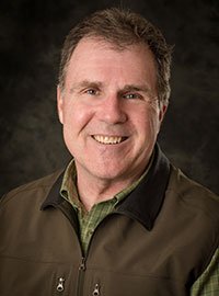
It is important to use the google reservation calendars now in case we need to get in touch with you.
Here are the instructions. https://www.mtu.edu/acmal/access/reservations/
It is important to use the google reservation calendars now in case we need to get in touch with you.
Here are the instructions. https://www.mtu.edu/acmal/access/reservations/
ACMAL lab managers are working within the group, with other Shared Facility directors and the VPR office to formulate lab procedures to be used during this period. You can expect to receive those plans by weeks end. Please feel free to email me any ideas you may have relative to using a facility shared with other faculty and graduate students.
Updated March 13, 2020.
Information for use of the facility during that time is included in this message.
Our overall message to you is please think ahead. If you know you will need help arrange that ahead of time.
Most of the ACMAL equipment/software is not compatible with remote operation or remote data analysis. In nearly all cases you will need to come to the facility to work. An exception is two XRD software programs. Ed Laitila can help you access them ealaitil@mtu.edu .
Use the calendar reservation system to book equipment time. XPS sessions can be scheduled by contacting Tim Leftwich trleftwi@mtu.edu. Avoid bringing multiple people to the lab with you. Work independently as much as possible. Special conditions apply for campus visitors who come to the labs.
ACMAL staff will do our best to accommodate training during the hiatus period, but it may not be available right away. Again, plan ahead.
Please wear gloves for all instrument use. ACMAL staff will be cleaning door knobs, sample holders, keyboards and mice, and knob set controls each day. You can cleanse surfaces yourself if you’d like with wipes that will be available next week.
Do not come to the labs sick. Please refer to the University information regarding self-identifying illness and self-isolation. If ACMAL staff are exposed to COVID-19 or infected, a unit in the facility may have to be closed.
It is important to note that this situation is changing daily and you should expect adjustments to current policies and procedures. I will send you updates as necessary.
Please let me know if you have any questions or concerns. Contact me if you need help. My phone number is 906-369-1875. I don’t use text very much so call me.
Job No: 20043
Department: Materials Science and Engineering
Work Type: Staff Full-time
Location: Main Campus (Houghton, MI)
Full Time / Part Time: Full Time
Categories: Management, Sciences
Michigan Tech seeks a director of its Applied Chemical and Morphological Analysis Laboratory (ACMAL). ACMAL is an interdisciplinary, university shared user facility that manages a suite of high-end structural characterization facilities including SEM (3 units), STEM (FEI Titan Themis), FIB, XPS, AFM, XRD, XRF, and associated sample preparation facilities. The director manages the access and operational procedures, coordinates maintenance, oversees the personnel needs of the facility, interfaces with the campus-wide user community, coordinates user training, and leads or participates in efforts to expand capabilities and use. The Director will have a staff appointment in the Materials Science and Engineering Department, who manages the facility in behalf of the university.
The ideal candidate will have extensive experience in the operation of electron-based imaging and analytical techniques in a research environment, be familiar with shared user-oriented facility model, demonstrated managerial skills, and a positive attitude compatible with providing efficient and high quality service to the university research community. A degree in the physical sciences, or engineering, or applied technology discipline is preferred.
Michigan Technological University is an Equal Opportunity Educational Institution/Equal Opportunity Employer that provides equal opportunity for all, including protected veterans and individuals with disabilities.
I will be out next week but I’ll mostly be in town. I could come in if needed. Aleister will be here working working 2 hours each day for coating samples and helping users. He plans to be here at midday.
The labs will be open all week between 8 – 5 and restricted users will work then. Unrestricted users have 24 hour access. Newly trained users, please work while Aleister is at work in case you have trouble.
Call Aleister or me if you have trouble. Aleister 520-576-3557. Owen 906-369-1875.
Thanks, Owen
We have made changes to the process of saturating the ESEM filament. Now we use Scan>Horizontal Line and monitor that line as the Filament slider is changed. Saturation occurs when the horizontal line stops increasing when the slide is increased. The point when it stops increasing is saturation, so you may overshoot and have to slide it back slightly. When you complete saturation return to Scan>Full Screen to stop the line. Click Filament Limit as you usually would. Any of you who learned to saturate the filament on the 6400 SEM will recognize this technique.
I want to show you how to do this so let me know when you’d want to see it done. It is really easy and there is no ambiguity when setting saturation like using the “football”. We will be making changes to the standard operating procedure.
This a a precise technique and the filament will last longer.
The ESEM is back online and ready to use.
I’m not certain what went wrong but I’m suspicious of the AZtec scan grabber. The AZtec software acquires an ESEM image by taking control of the ESEM scan hardware. When the scan is complete it should release the ESEM hardware. I have seen a few times that it doesn’t release. If you have trouble seeing a scanning ESEM image after acquiring with AZtec try to shut down the AZtec software and see if the scan returns. If it still down’t return contact me and I’ll take a look at it.
The ESEM is down with a computer problem. Don’t try to use it. I’ll
get it back online Monday morning and send out an update.
Chathura de Alwis (Chemistry), Timothy R. Leftwich (MSE), Pinaki Mukherjee (MSE), Alex Denofrea (Chemistry) and Kathryn A. Perrine (Chemistry) published a paper titled “Spontaneous selective deposition of iron oxide nanoparticles on graphite as model catalysts” in Nanoscale Advances in 2019.
DOI: 10.1039/c9na00472f
Iron oxide nanomaterials participate in redox processes that give them ideal properties for their use as earth-abundant catalysts. Fabricating nanocatalysts for such applications requires detailed knowledge of the deposition and growth. We report the spontaneous deposition of iron oxide nanoparticles on HOPG in defect areas and on step edges from a metal precursor solution.
Various defects were created on the highly oriented pyrolytic graphite (HOPG) surface using either argon (Ar+) sputtering or a focused ion beam (FIB) to provide defects for nucleation sites. A Hitachi 2000 A FIB instrument was used to create tailored arrays of defects on HOPG using a Ga+ beam.
The sputter rate was calculated using the amount of materials removed, by recording a height profile of 1 nm using atomic force microscopy (AFM) and the time to sputter the pattern.
All the samples were imaged using a Hitachi S-4700 cold field emission high resolution field emission scanning electron microscopy (FE-SEM) instrument.
X-ray photoelectron spectroscopy (XPS) was performed using a PHI 5800 to analyze the elemental composition and oxidation state of surface species of the iron oxide nanoparticles grown on the HOPG surface.
Scanning transmission electron microscopy (STEM) imaging and energy dispersive X-ray spectroscopy (EDS) mapping were used to measure the phase and composition of iron oxide nanoparticles after annealing and to confirm if the deposition was preferential at the defect sites of graphite. A FEI Titan Themis aberration corrected scanning transmission electron microscope was used to obtain atomically resolved electron images and EDS maps of the iron oxide nanoparticles on the graphene coated TEM grid. The microscope was operated at 200 kV using a point resolution of the aberration corrected STEM mode of 0.08 nm. The microscope was equipped with a SuperXâ„¢ X-ray detector, which is composed of 4 detectors for fast X-ray mapping in STEM mode. The EDS mapping of the sample was performed on specific particles with an average beam current of 100 pA.
Equipment for obtaining the AFM images in this project was provided by NSF CHE #1725818. The electron microscopy research was performed at the Applied Chemical and Morphological Analysis Laboratory, at Michigan Technological University. The electron microscopy facility is supported by NSF MRI 1429232. We acknowledge the Michigan Tech REF-RS fund for support of this work and the David J. and Valeria Pruett Graduate Research Fellowship. We acknowledge the Applied Chemical and Morphological Analysis Laboratory (ACMAL) for staff assistance and use of facilities.
de Alwis, C., Leftwich, T., Mukherjee, P., Denofre, A., & Perrine, K. (2019). Spontaneous selective deposition of iron oxide nanoparticles on graphite as model catalysts. Nanoscale Advances, 1(12), 4729-4744.
http://doi.org/10.1039/C9NA00472F
Retrieved from: https://digitalcommons.mtu.edu/michigantech-p/1246
Pinaki is out with an illness likely the remainder of this week. He will be rescheduling appointments.
Liz Miller will be out of town for 6 weeks beginning next week. Aleister and I will try to pick up the work she was doing during her absence. One area that he and I cannot assume is EBSD. We have asked someone to help us with that but the response will be slowed. So, plan your EBSD work ahead of time – don’t wait until the last minute or your work will be delayed.
Liz will return to work on April 6th.