Category: Research

Sangyoon Han Wins Best Poster Award for Cell Adhesion Research

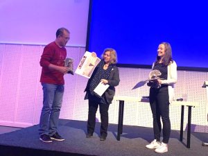
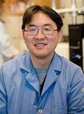
Sangyoon Han Accepts Award
Sangyoon Han (left) accepts the poster prize. Image from Alessandra Cambi.

Sangyoon Han (biomedical engineering) was awarded the Dutch Society of Cell Biology’s Best Poster Award for his work on cell adhesion and migration in inflammation and cancer.

The title of his poster is “Talin-vinculin pre-complex formation dictates maturation of nascent adhesions by accelerated force transmission and vinculin recruitment.”

The 6th ZOO Meeting took place at Blijdorp Zoo, Rotterdam, The Netherlands, May 15-18, 2019.

The ZOO meeting series has become a landmark event in the field of cell adhesion and migration due to unique theme selection, high scientific profile with excellent speakers and limited number of attendees.

Invited Review of Multifunctional Biomedical Adhesives

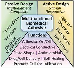
Multifunctional Biomedical Adhesives passive design active design functionsAdvanced Health Materials published the article “Multifunctional Biomedical Adhesives,” by Bruce Lee, Rupak Rajachar, Rattapol Pinnaratip, Saleh Akram Bhuiyan, and Kaylee Meyers (Biomed). This article is an invited review that described recent advances in designing multifunctional bioadhesives for various biomedical applications.

https://doi.org/10.1002/adhm.201801568

This review discusses strategies for engineering multifunctional biomedical adhesives, which involve two general approaches: passive and active design. Passive adhesives contain inherent structural elements that can carry out additional functions without external influences, whereas active adhesives are designed to respond to environmental changes. These adhesives exhibit new functionality such as antimicrobial properties, self‐healing ability, and the capacity to release drugs.

Wireless Sensor Project Funding for Keat Ghee Ong

Keat Ghee Ong
Keat Ghee Ong

Keat Ghee Ong (BioMed) is the principal investigator on a project that has received a $14,000 research and development grant from the University of Oregon.

The project is titled “Implementation of a Wireless Sensor System for Monitoring Mechanical Loadings at the Internal Fixation Plates of Rats with Segmented Bone Defects.”

This is the first year of a potential three year grant totaling $54,000.

By Sponsored Programs.

DHH-NIH Funding for Jeremy Goldman

Jeremy Goldman (Bio Med) is the principal investigator on a project that has received a $45,790 research and development grant from the Department of Health and Human Services-National Institutes of Health.

Jaroslaw Drelich (MSE) is the Co-PI on the project “Inhibition of Neointimal Hyperplasia by Zinc-Based Biodegradable Arterial Devices.”

This is the first year of a potential two-year project.

By Sponsored Programs.

Jeremy Goldman
Jeremy Goldman
Jarek Drelich
Jarek Drelich

DHH-NIH Funding for Feng Zhao

Feng Zhao (Bio Med) is the principal investigator on a project that has received a $459,000 research and development grant from Department of Health and Human Services – National Institutes of Health. Jeremy Goldman (Bio Med) is Co-PI on the project, “Development of Off-the-Shelf Completely Biological Small-Diameter Blood Vessel with Human Stem Cells.”

The is the first year of a potential three-year project.

Feng Zhao
Feng Zhao
Jeremy Goldman
Jeremy Goldman

Biomedical Engineering Researchers Attend Adhesion Society Meeting

Adhesion Society Annual Meeting location photoBruce Lee (BioMed), Rupak Rajachar (BioMed), Ameya Narkar, Ariana Tyo and Saleh Akram attended the 42 Annual Meeting of the Adhesion Society in Hilton Head, South Carolina.

Lee served as the chair of the Bioadhesion Division within the Adhesion Society and was one of the organizers in the meeting. Rajachar chaired two sessions entitled “Interfaces in Pharmaceutical Sciences” and “Bioadhesive Chemistry.”

Narkar gave an oral presentation entitled “Evaluating Rapid Switching and Reversible Adhesion of Adhesive Hydrogel-Coated PDMS Micropillars,” a project directed by Lee. Narkar also co-chaired a session entitled “Bioadhesive Chemistry.”

Tyo gave an oral presentation entitled “Optimizing of Two-Step Adhesive Coating for the Mitigation of Field Associated Infection in Cetacean Satellite Telemetry Tags,” a project directed by Rajachar.

Akram gave an oral presentation entitled “Controlling Redox Reaction of Conductive Smart Catechol Adhesive using Electrochemistry,” a project directed by Lee.

The meeting took place February 17-20, 2019.

Suspicious Mammograms: Taking the Guesswork Out of Elastographic Ultrasounds

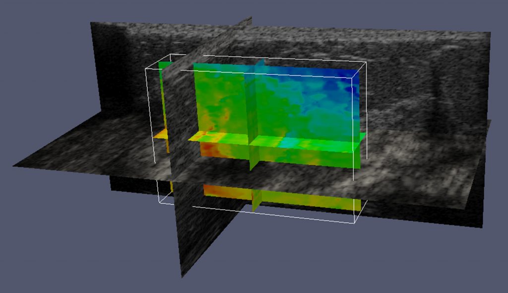
Jingfeng Jiang uses a graphics processing unit (GPU) to perform advanced processing of raw ultrasound data
Jingfeng Jiang uses a graphics processing unit (GPU) to perform advanced processing of raw ultrasound data, to help radiologists better evaluate suspicious mammograms.

Jingfeng Jiang is the principal investigator on a project that has received a $450,187 research and development grant from the National Institutes of Health, “Elastography-Based Analytics for Benign and Malignant Breast Disease.”

Jingfeng Jiang
Assoc. Professor Jingfeng Jiang, Michigan Tech

Ultrasound elastography is used to pinpoint possible tumors and differentiate malignant, cancerous growths from benign lesions throughout the body, including in the breast. “Cancer tissues are stiff, and aggressively change their surroundings,” says Jiang, an associate professor of biomedical engineering at Michigan Tech.

“Ultrasound elastography uses imaging to measure the stiffness of tissue. Depending on who does the reading, the accuracy can vary from 95 percent to 40 percent,” Jiang says. “Forty percent is very bad—you get 50 percent when you toss a coin. In part, the problem is that ultrasound elastography is a relatively new modality.”

Ultrasound elastography could be an excellent screening tool for women who have suspicious mammograms, but only if the results are properly interpreted. Jiang’s research team, along with Zhengfu Xu, assistant professor of mathematical sciences, will use their graphics processing unit (GPU) to perform advanced processing of raw ultrasound data so physicians can use that information in their clinical workflow. “Mainly, radiologists will use our software together with ultrasound elastography and ultrasound for diagnosis,” says Jiang. “Our goal is to greatly reduce the guesswork.”

Biomedical Research Featured in SME’s Humans of Manufacturing

Humans of ManufacturingResearch by biomedical engineers at Michigan Tech was featured in the article “SME’s Humans of Manufacturing — Developing Hearts,” in Additive Manufacturing. The article focuses on Dr. Martin Bocks’ efforts to solve cardiology problems in small children.

Developing Hearts

Dr. Martin Bocks Seeks to Solve Big Cardiology Problems in Small Children

“Right now, we use stents that are FDA approved for adult indications such as coronary or peripheral vascular disease, or stents for the biliary tract,” said Dr. Martin Bocks, a pediatric cardiology specialist at the Cleveland Ohio’s UH Rainbow Babies & Children’s Hospital.

Working with biomedical engineers from Michigan Tech University and Case Western Reserve University, he has developed a new type of stent made of a special zinc alloy that safely degrades over time and is gradually taken up by the body. It is not permanent like those made of Nitinol, stainless steel, or other bare metals, but shares many of the same properties.

Dr. Bocks explains that the manufacturing process is much the same as any other metal stent, except that the zinc is much harder to work with. “We start with the raw material provided by the team at Michigan Tech, have it cast into rods, and extruded into tubes or a cannulae, which are then laser cut according to our stent design. It’s electro-polished, crimped onto a balloon, and delivered. So far, it looks like an excellent alternative for pediatric patients.”

Read more at SME, Smart Manufacturing Experience, by SME Media.