Category: Biomedical Engineering

Bruce Lee: Smart Biomaterials Inspired by Mussel Chemistry

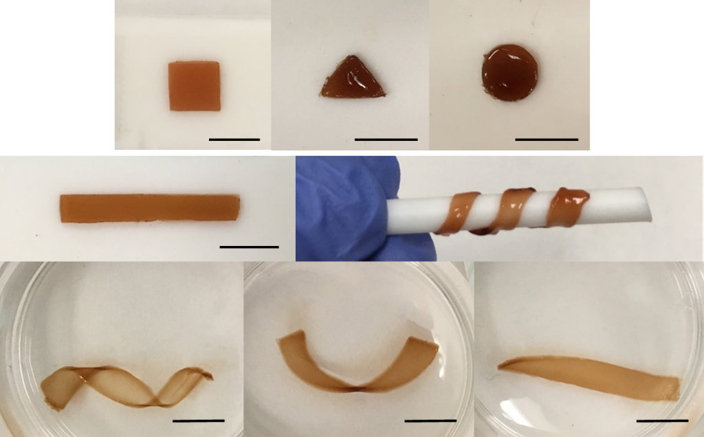
Self-healable and moldable nanocomposite gel as fit-to-shape sealant.
Bruce Lee exploits the ability of a Dopa, a unique catechol-based amino acid found in mussel adhesive proteins, in a new fit-to-shape sealant. It initially exhibits the ability to be remolded and adhered to the convex contour of a tissue surface. With time, the hydrogel is fixed in its new shape.

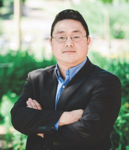
Bruce Lee, an associate professor of biomedical engineering at Michigan Tech, focuses on smart adhesives and biomaterials inspired by nature. More specifically, the natural glues made by mussels that anchor them to rocks, boats and docks. His past work on hydrogels and tissue adhesives led him to look more closely at what makes these adhesives work underwater—and how people could use them.

Lee’s research team exploits the ability of Dopa, a unique catechol-based amino acid found in mussel adhesive proteins. He currently has three active, federally funded research projects.
Bruce Lee, Associate Professor of Biomedical Engineering, Michigan Tech

Smart Adhesive

As a participant in the Office of Naval Research (ONR) Young Investigator Program, Lee delves into not only what makes mussels sticky but also how to reverse that adhesion with an electrical charge. The YIP grant is awarded to scientists and engineers with exceptional promise for producing creative, state-of-the-art research that appears likely to advance naval capabilities. “There is no smart adhesive out there that can perform underwater,” he says. “The chemistry that we can incorporate into the adhesive, causing it to reversibly bond and de-bond, is quite new.”
Fit-to-Shape Sealant
Lee is also designing an injectable sealant and bioadhesive, funded by the National Institutes of Health (NIH). Lee and his research team developed a moldable nanocomposite hydrogel. “This material initially exhibits the ability to be remolded and adhered to the convex contour of a tissue surface,” says Lee. “With time, the hydrogel is fixed in its new shape and functions as a fit-to-shape sealant.” Their hydrogel uses no cytotoxic crosslinking reagent, and needs no mixing tip for mixing precursor solutions. It also demonstrates burst pressure potentially suited for sealing renal vein and even intestinal anastomosis. “One very valuable quality of this synthetic glue is its versatility,” adds Lee. “We can change the chemistry to make it as rigid or flexible as we need — while still maintaining its overall strength and durability.”
Smart Antimicrobial Microparticles
Lee just received new funding from the Office of the Assistant Secretary of Defense for Health (OASDH) to design smart microparticles from mussel-derived catechol. “The particles are pathogenic and able to promote healing,” Lee says. “Simply hydrating the microparticles in water causes them to generate hydrogen peroxide that can kill bacteria and inactivate viruses. This material can potentially function as a lightweight and portable disinfectant for a wide range of applications.”
Visit Lee’s research group online to learn more about their bio-inspired approach to the design of advanced functional materials.

Systems Engineering Research Center Supports Undergraduate Student Projects

SERC

Joseph Thompson, Zachary Fredin and Richard Berkey of the Pavlis Honors College will receive $60,000 in undergraduate student project funding from the Systems Engineering Research Center (SERC). SERC is a University Affiliated Research Center of the Department of Defense that collaborates with 22 universities across the United States to leverage the expertise of senior lead researchers. SERC represents a broad community of systems engineering researchers whose depth of knowledge spans a wide range of diverse interests and industries.

The initial 12 projects, funded through SERC, will provide students in biomedical engineering, electrical engineering and five different Enterprises with valuable hands-on experience serving Naval Systems Warfare, Army, Air Force Special Operations, Air Force Research Laboratory, Marine Corps Special Operations Command, United States Coast Guard and United States Special Operations Forces.

Inaugural project work will take place throughout the 2018-19 academic year.

By the Pavlis Honors College.

May the Force Be with You: Sangyoon Han Brings Mechanobiology to Michigan Tech

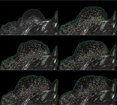
Tracked adhesion population classified with Machine Learning. Sangyoon Han uses images like these to measure and compare force behavior.
Sangyoon Han uses images of live cells to measure and compare force behavior. Pictured here: tracked adhesion population, classified with Machine Learning.

Cancer cell metastasis. Stem cell differentiation. Atherosclerosis. All are strong mechanotransduction-related physiological and pathophysiological events. Just how do cells transduce mechanical force into biochemical signals? 

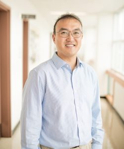
Assistant Professor Sangyoon Han, Department of Biomedical Engineering, Michigan Tech
Assistant Professor Sangyoon Han, Department of Biomedical Engineering, Michigan Tech.

“Cells are sensitive to mechanical forces outside the cell membrane,” says Sangyoon Han, who joined the Department of Biomedical Engineering at Michigan Tech as an assistant professor last fall. At their basal surface, however, cells are interfacing with something called the extracellular matrix (ECM), which supports the cell not only chemically but also mechanically.”

“Over the past 20 years, it has been revealed that the rigidity of the extracellular matrix can greatly influence the physiology and pathology of cells and tissues, including differentiation, survival, proliferation, altered drug response, and tumor progression,” adds Han. “In the case of a tumor, an increase in tissue stiffness—without any changes in genetic information and chemical environment—can cause tumor progression. There is also an evidence showing that cancer-targeting drugs do not work when cancer cells are highly contractile in a very tensed environment,” he says.

To investigate this, Han and his team established experimental and computational frameworks for force measurement and adhesion dynamics quantification. “We apply these frameworks, with cutting-edge computer vision techniques, on live-cell microscope images to find out the fundamental mechanisms underlying mechanosensation in normal cells, as well as the biomechanical signature in diseased cells whose signaling has gone awry.”

Han measures the force a cell transmits to the environment with traction force microscopy. “The force sensor, referred to as a focal adhesion, consists of a special receptor across the membrane and over 100 cytoskeletal adaptor proteins. These focal adhesion proteins have redundant and diverse roles in signaling and structural development of the adhesion,” he explains.

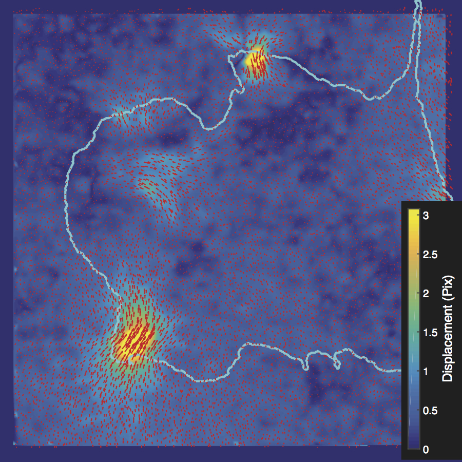
L to R: adhesion segmentation, displacement map, and tracking map. Photo credit: Sangyoon Han, Michigan Tech
L to R: adhesion segmentation, displacement map, and traction force map. Photo credit: Sangyoon Han, Michigan Tech

Using high-resolution imaging of living cells on a soft substrate, Han captures gel deformation and force-sensing protein trajectories at the same time. Han’s novel force-reconstruction software converts the measured gel deformation into a force map over a cell footprint. Using time-series data extracted from the image data, he monitors feedback between the cellular structure and its mechanical forces.

Han shares his Matlab-based, open-source software with the mechanobiology community. In his Mechanobiology Lab at Michigan Tech, Han is also building a physical device using bioMEMS for active force application to cells and tissue. “I firmly believe that engineers can make significant contributions to not only the biomedical industry, but also fundamental biological science.”

Before coming to Michigan Tech, Han was a postdoctoral researcher at the Harvard Medical School Lab of Computational Cell Biology, as well as the University of Texas Southwestern Medical Center. He earned a PhD in Mechanical Engineering at University of Washington in the area of cell mechanics, multiphysics modeling, and bioMEMS, and BS and MS in Mechanical Engineering at Seoul National University.

Color-coded map of deformation of a gel, quantified using the fluorescent beads. Photo Credit: Sangyoon Han, Michigan Tech


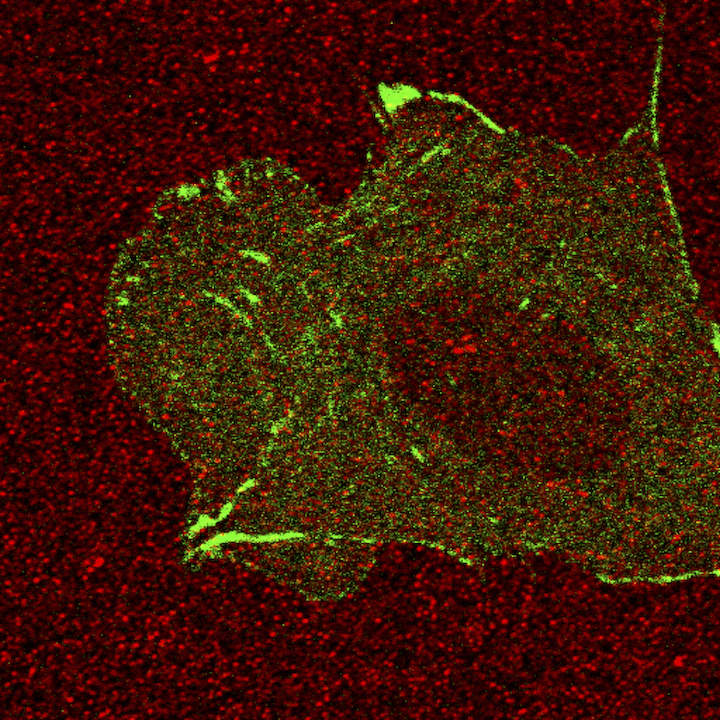
Red spots are the fluorescent beads coated on top of the gel, which we use to quantify the deformation of the gel. Green signal is the paxillin, one of the focal adhesion proteins of a Chinese Hamster ovary cell. Photo credit: Sangyoon Han, Michigan Tech
“Red spots are the fluorescent beads coated on top of the gel, which we use to quantify the deformation of the gel,” explains Sangyoon Han, assistant professor of biomedical engineering at Michigan Technological University. “Green signal is the paxillin, one of the focal adhesion proteins of a Chinese Hamster ovary cell.”

Graduate School Announces Fall 2018 Award Recipients

Engineering Grad Students working in the lab

The Graduate School announced the Summer and Fall 2018 award recipients. The following are award recipients in engineering graduate programs:

Doctoral Finishing Fellowship Award

Ulises Gracida Alvarez, Chemical Engineering
Sanaz Habibi, Chemical Engineering
Long Zhang, Chemical Engineering
Shuaidong Zhao, Civil Engineering
Jingyuan Wang, Electrical Engineering
Zhimin Song, Environmental Engineering
Priscilla Addison, Geological Engineering
Hans Lechner, Geology
Huaguang Wang, Materials Science and Engineering
Shadi DaraniMechanical Engineering-Engineering Mechanics
Soroush Sepahyar, Mechanical Engineering-Engineering Mechanics

Portage Health Foundation Graduate Assistantship

Anindya Majumdar, Biomedical Engineering
David Rosen, Biomedical Engineering

Dean’s Award for Outstanding Scholar

Shuaidong Zhao (Civil and Environmental Engineering PhD)
Priscilla Addison(Geological Engineering PhD)
Sampath Kumar Reddy Boyapally (Mechanical Engineering MS)
Rahul Jitendra Thakkar (Mechanical Engineering MS)
Nikhil Appasaheb Shinde(Mechanical Engineering MS)
Mitchel Timm (Mechanical Engineering MS)
Xinyu Ye (Environmental Engineering PhD)

Dean’s Award for Outstanding Graduate Student Teaching

Dongdong Ge (Civil and Environmental Engineering PhD)
Mohammadhossein Sadeghiamirshahidi (Civil and Environmental Engineering PhD)
Aaron Krieg (Chemical Engineering PhD)
Brandi Petryk (Geology MS)
Christa Meingast (Environmental Engineering PhD)
Luke Jurmu (Mechanical Engineering-Engineering Mechanics PhD)
Mingyang Li (Mechanical Engineering-Engineering Mechanics PhD)

North Macomb Students Attend Women in Engineering Program

Women in EngineeringA trio of local students recently had a chance to explore an array of engineering careers through Michigan Technological University’s Women in Engineering program.

The Women in Engineering program is a weeklong look at engineering careers in areas such as mechanical, computer, environmental, electrical, biomedical, civil, geological and materials engineering, school officials said in a news release.

Students accepted into the program received a scholarship that covered room and board, tuition and supplies.

Read more at The Voice, by Emily Pauling.

Narkar and Lee on pH-Responsive, Reversible Adhesion

Ameya Narkar and Bruce Lee (Biomed) published “Incorporation of Anionic Monomer to Tune the Reversible Catechol-Boronate Complex for pH Responsive, Reversible Adhesion,” in Langmuir (ACS Publications).

DOI: 10.1021/acs.langmuir.8b00373

Bruce P. Lee is an associate professor in the Department of Biomedical Engineering. Ameya Narkar is a PhD student in the biomedical engineering program.

Ameya Narkar
Ameya Narkar
Bruce P. Lee
Bruce P. Lee

Analyzing the Behavior of Light in New Zealand

Mitch Kirby at Westland Tai Poutini National Park, New Zealand
Mitch Kirby at Westland Tai Poutini National Park, New Zealand

The natural beauty and easy access to both snowboarding and surfing first attracted Mitch Kirby to New Zealand—that, and the legendary fly fishing. Kirkby was a sophomore majoring in biomedical engineering at Michigan Technological University when he received a Whitaker International Student Fellowship at the University of Otago in Dunedin, New Zealand.

“One of my professors at Michigan Tech, Dr. Sean Kirkpatrick, told me about the Biophotonics and Biomedical Imaging Research Group at the University of Otago. As I learned more about New Zealand, everything sort of seemed to line up.”

Kirby worked with a group focused on light/tissue interaction. “As light propagates through biological tissue, the light waves exhibit different behavior based on the internal characteristics of the tissue,” Kirby explains. “Ultimately the goal of the project was to gather enough experimental data on the different light-tissue interactions so that down the road it would be possible to use a light-emitting device to make medical diagnostic decisions non-invasively. While the project was in the early stages, most of my time in the lab was spent lining up the different lenses and filters for the experiments with elliptically-polarized light. Later we began writing code on MATLAB and analyzing the behavior of the light.”

Kirby’s everyday life in Dunedin involved getting up very early, completing schoolwork and attending classes. After spending a few hours in the lab, he would finish up for the day around 3 pm. If the waves were good, he would surf. If not, he would explore the countryside. During the weekends, he traveled with a small group of friends to different locations throughout New Zealand. Trips usually involved snowboarding, backpacking, and just general adventuring.

Mitch Kirby crosses the Copeland Valley in Westland Tai Poutini National Park, New Zealand
Mitch Kirby crosses the Copeland Valley in Westland Tai Poutini National Park, New Zealand

“Spending time overseas definitely opened my eyes to the ability of a college education to take you places,” says Kirby. “Traveling and living abroad while studying and working in the lab showed me that it is possible to mix work and play so that each day is an enjoyable one,” he adds. “I also enjoyed the excitement of working on a research project that could potentially change the way many medical diagnoses are made. There is a great deal of potential in the continued advancement of biomedical optics. My ultimate goal is to develop new technologies through academic research.”

Working with people in the lab from different backgrounds was a high point for Kirby as well. “Everyone had something unique to bring to the table, particularly because we all came from different countries and cultures.”

Michigan Tech biomedical engineering student Mitch Kirby surfs in New Zealand. "Traveling and living abroad while studying and working in the lab showed me that it is possible to mix work and play so that each day is an enjoyable one.”
Michigan Tech biomedical engineering student Mitch Kirby surfs in New Zealand. “Traveling and living abroad while studying and working in the lab showed me that it is possible to mix work and play so that each day is an enjoyable one.”

Once back in Michigan Tech, Kirby returned to the optics lab at Michigan Tech to investigate Optical Coherence Tomography as an undergraduate researcher.

Mitch Kirby earned a BS in Biomedical Engineering from Michigan Tech in 2016. The same year he received a National Science Foundation Graduate Research Fellowship and is now a doctoral student in Bioengineering and Biomedical Engineering at the University of Washington in Seattle.

Four New Biomedical Engineering Minors for Fall 2018

Biomedical Engineering MinorsBiomedical engineering is a rapidly growing and evolving field. The need to have a well trained workforce with the ability to integrate life sciences, engineering, and the practices of modern medicine is a pressing issue.

The Department of Biomedical Engineering is offering four new minors related to biomedical engineering beginning Fall 2018:

  • Biomaterials Engineering
  • Biomedical Engineering
  • Medical Devices and Instrumentation
  • Tissue and Stem Cell Engineering

The minor programs will help to prepare students for careers in the medical device or related industry sectors. They may pursue graduate study at the interface of life science and engineering. The minors also help prepare students for professional careers, such as medicine, dentistry, physical therapy, or occupational therapy.

Michigan Tech invites students from all disciplines to learn the fundamental concepts of biomedical engineering. The minors are structured in such a manner that they are accessible to a broad range of majors, such as materials science and engineering, chemical engineering, electrical engineering, general engineering, and mechanical engineering. Science majors can take these minors if the pre-requisite math and engineering courses are met.

Students will broadly understand key concepts and principles of biomedical engineering. They will develop the beginnings of an understanding of how the life sciences and other engineering disciplines can be integrated to solve biomedical engineering problems.

Research Excellence Fund Awards Announced for 2018

Jeremy Bos in the labThe Vice President for Research Office announced the 2018 Research Excellence Fund (REF) awards and thanked the volunteer review committees, as well as the deans and department chairs, for their time spent on this important internal research award process. The awardees in the College of Engineering are listed below:

Infrastructure Enhancement (IE) Grants

Portage Health Foundation (PHF) Infrastructure Enhancement (IE) Grants

  • Jingfeng Jiang “JJ” (BME/LSTI) – Electromechanical Biomechanical testing apparatus (ACUMEN [3KN systems])

Research Seed (RS) Grants

Portage Health Foundation (PHF) Mid-Career (MC)

BME Researchers Review Tissue Engineered Vascular Graft Advancements

TEVG Diagram of blood flow and components
Developing a biocompatible blood-contacting surface remains a major challenge for tissue engineered vascular grafts (TEVGs). This paper reviews the current state of TEVGs with an emphasis on the blood-contacting surface, which includes general vascular physiology and developmental challenges, materials currently employed in TEVGs, and strategies to modify blood-contacting surfaces to resist thrombosis and control cellular recruitment.

Tissue engineered vascular grafts (TEVGs) are beginning to achieve clinical success and hold promise as a source of grafting material when donor grafts are unsuitable or unavailable.

Daniel Radke, Wenkai Jia, Dhavan Sharma, Kemin Fena, Guifang Wang, Jeremy Goldman, and Feng Zhao have a review accepted in Advanced Healthcare Materials. The article “Tissue Engineering at the Blood-Contacting Surface: A Review of Challenges and Strategies in Vascular Graft Development” is an invited review which is a follow-up to a previous research publication: “Aligned nanofibrous cell-derived extracellular matrix for anisotropic vascular graft construction,” Advanced Healthcare Materials. 2017; 6:1601333 (1-6).

This is a timely and comprehensive review article that references extensive publications. It covers significant technological advances regarding tissue engineered vascular grafts for cardiac disease treatment. The review focuses on the challenge of developing a biocompatible blood-contacting surface.

The research group includes faculty, graduate, and undergraduate students in the department.